Magnetic Force Microscopy
AFM is a versatile measurement technique that allows investigation of various surface properties in addition to the standard topography mapping. One of the secondary techniques is Magnetic Force Microscopy (MFM) which enables high-resolution imaging of the magnetic properties of different materials. By using a probe with a magnetic coating on the tip side of the cantilever, the AFM system is capable of qualitatively measuring the magnetic field gradients above the sample surface.
The measurements can be performed in either static or dynamic mode. In static mode, the deflection of a nonoscillating cantilever above the surface due to magnetic interactions is detected and in dynamic mode - the amplitude or the phase of the cantilever oscillation (Fig. 1). Dynamic mode MFM is usually preferred for its higher sensitivity.
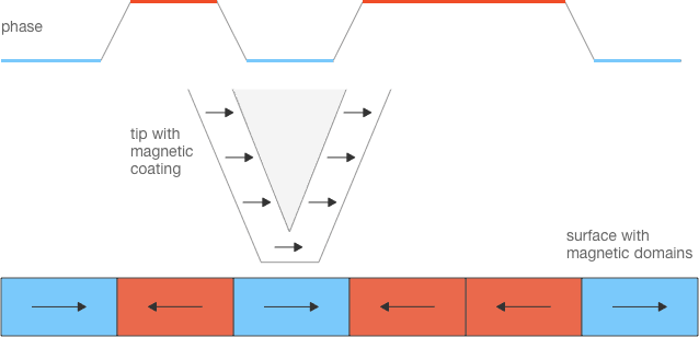
MFM measurements are qualitative because of the complex magnetization of the tip’s magnetic coating and the tip–sample interactions which affect both the tip and the sample. Most commercially available probes offer hard magnetic coatings whose purpose is to retain their magnetic polarization orientation as long as possible. However, there are a few models with soft magnetic coatings which are intended to bring less disturbance when imaging soft magnetic samples.
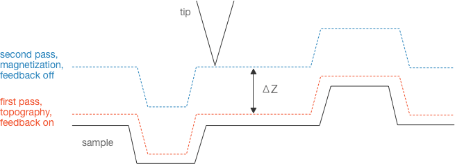
In order to separate the magnetic and the topographic information, a special two-pass imaging mode is commonly used (Fig. 2). During the first pass the probe tracks the topography of a single scan line and then retracts from the surface. During the second pass the probe follows the topography at a predefined constant height ΔZ (usually several tens to several hundred nanometers) with the feedback disabled, detecting only the magnetic interactions
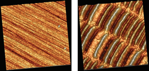
Typical applications of MFM are imaging magnetic bits of magnetic data storage devices and domains on magnetic materials. The images on Fig. 3 are obtained from a measurement on a hard disk drive (HDD) platter surface. The first image, consisting of all the first pass scan lines, shows the surface topography. The second image, comprising the second pass scan lines, shows the variations of the magnetic field. The high and low areas in the magnetic image are regions with different orientation of the magnetic dipoles that store binary 1’s and 0’s.
At present, MFM allows typical resolutions below 50nm. Previously, such resolutions were only achievable by electron microscope techniques, such as Spin-Polarized Low-Energy Electron Microscopy (SPLEEM). MFM offers the advantage of little or no sample preparation needed and the possibility to obtain real 3-dimensional images.
For more information on BudgetSensors MFM probes, visit www.budgetsensors.com/magnetic-afm-probes