

BudgetSensonsors AFM Calibration NanogridMon Apr 27 2020


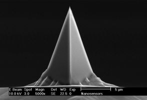
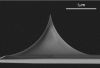
Discover how the wear behavior of nanotools high densitiy carbon CDR50-EBD AFM probesThu Apr 23 2020
Discover how the wear behavior of nanotools high densitiy carbon CDR50-EBD AFM probes with controlled 50 nm diameter compares to that of standard CDR silicon probes.


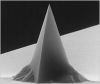
3D‐Printed Scanning‐Probe MicroscopesMon Apr 13 2020
3D‐Printed Scanning‐Probe Microscopes with Integrated Optical Actuation and Read‐Out'
Blow the dust off your good ol' 3D printer and print yourself an Atomic Force Microscope! : )

The Microscope That Uses Quantum Physics to Trace AtomsFri Apr 10 2020


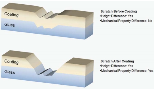
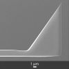
MikroMasch® HQ:NSC36 series AFM probesMon Mar 23 2020
Defect recognition on coating layer with MikroMasch® HQ:NSC36 series AFM probes using PinPoint nanomechanical mode


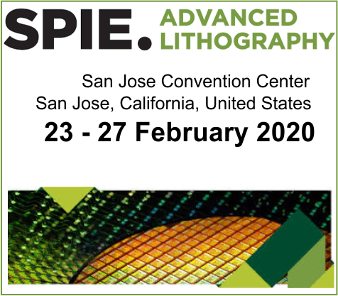
Nanotools is attending the SPIE Advanced Lithography in CaliforniaFri Feb 21 2020
nanotools is attending the SPIE Advanced Lithography in California.
The technical program of the primary global #lithography event will focus
If you would like to meet with nanotools in San Jose then please click on the contact link in the nanotools blog https://www.nanotools.com/…/connecting-with-partners-and…


It’s the last day @BiophysicalSoc Meeting 2020 exhibit in San DiegoTue Feb 18 2020
It’s the last day @BiophysicalSoc Meeting 2020 exhibit in San Diego and it’s the last chance to visit @NanoAndMore USA booth no. 818 from 10 am – 4 p.m. to pick up your free sample of NANOSENSORS™ uniqprobe qp-BioT #AFMprobes and to find out about all the other kinds of #AFMtips that NanoAndMore USA offers for #AtomicForceMicroscopy applications in #biology, #biophysics, #molecularbiology, #lifesciences etc. etc. … https://www.nanoandmore.com/Life-Science-Biological-Soft…
We’re looking forward to welcoming you! #bps2020
Highlights


AFM Probe Focus


qp-HBC
uniqprobe™ - HeartBeat Cantilever for ScanAsyst®** and Peak Force Tapping™**
Coating:
Reflective Aluminum
Tip Shape: Circular symmetric
Tip Shape: Circular symmetric
AFM Cantilever:
F
60 kHz
C
0.5 N/m
L
115 µm
ATEC-NC
Tapping Mode AFM Probe with REAL Tip Visibility
Coating:
none
Tip Shape: Visible
Tip Shape: Visible
AFM Cantilever:
F
335 kHz
C
45 N/m
L
160 µm
TESPA
Standard Tapping Mode AFM Probe
Coating:
Reflective Aluminum
Tip Shape: Standard
Tip Shape: Standard
AFM Cantilever:
F
320 kHz
C
42 N/m
L
125 µm
OTESPA
Standard Tapping Mode AFM Probe with AFM Tip at the Very End of the AFM Cantilever
Coating:
Reflective Aluminum
Tip Shape: Optimized Positioning
Tip Shape: Optimized Positioning
AFM Cantilever:
F
300 kHz
C
26 N/m
L
160 µm