
 in 22-27 Aug.2022")
2022年8月22日~27日に金沢大学ナノ生命科学研究所(WPI-NanoLSI) において第 10回Bio- SPM夏の学校が開催されましたWed Aug 31 2022
2022年8月22日~27日に金沢大学ナノ生命科学研究所(WPI-NanoLSI) において第
10回Bio- SPM夏の学校が開催されました。https://nanolsi.kanazawa-u.ac.jp/…/applicatio…/summerschool/
㈱NanoAndMoreジャパンも本活動を支援させていただきました。
最終日には開催期間中の研究成果から、4名の研究者に対し奨励賞が授与され、
加えてNanoAndMoreジャパン https://www.nanoandmore.jp/ から副賞が渡されました。
NanoAndMore Japan proudly sponsored this year's Bio-SPM Summer SchoolWed Aug 31 2022
NanoAndMore Japan proudly sponsored this year's Bio-SPM Summer School at
Kanazawa University (WPI-NanoLSI) in 22-27 Aug.2022.
https://nanolsi.kanazawa-u.ac.jp/…/applicatio…/summerschool/
https://nanolsi.kanazawa-u.ac.jp/…/applicatio…/summerschool/
The 4 winners of the best image contest got a supplementary prize from NanoAndMore Japan. https://www.nanoandmore.jp/
Congratulations!


MikroMasch® platinum coated HQ:NSC18/Pt and NANOSENSORS™ conductive diamond coated CDT-CONTR AFM probes are used in this work on time dependent electrical contact resistance.Wed Aug 24 2022
Study on contact aging and its effect on electrical resistance in Conductive AFM measurements with the help of our MikroMasch® HQ:NSC18/Pt AFM probes


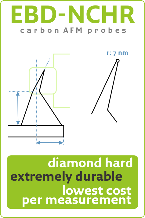
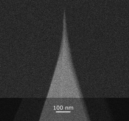
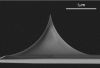
Metrology Super Sharp (MSS): Heavy-Duty Carbon Tips for Very Narrow HAR TrenchesMon Aug 22 2022
Blue Line product highlight:
MSS 4-Star: A durable depth metrology solution for consistent bottom access of finest features
MSS 4-Star: A durable depth metrology solution for consistent bottom access of finest features
- Controlled shape:
Precisely controlled length of 100 nm and width of 10 nm measured at 50 nm from apex - Controlled orientation: 12° or 3°
Tilt compensation within ±1° or ±0.5° for enhanced access capabilities to bottom trench features - Optimized cantilevers:
Standard with with k: 2.8 N/m, f: 75 kHz or k: 40 N/m, f: 320 kHz
Other cantilevers available upon request - Diamond-like hardness and durability:
Unmatched diamond-like carbon wear resistance for consistent performance and reduced cost per measurement - Delivered with TrueDimensions™:
Datasheet for every single probe available online 24/7 via QR code


ElectriMulti75-G and ElectriTap300-G AFM probes perform measurements in KPFM modeThu Aug 11 2022
Our ElectriMulti75-G and ElectriTap300-G AFM probes perform measurements in KPFM mode to study corrosion at the buried interface of organic films and Al alloy.


NANOSCALE NONCOLLINEAR SPIN TEXTURES IN THIN FILMS OF A D2D HEUSLER COMPOUNDTue Aug 09 2022
Nanoscale Noncollinear Spin Textures in Thin Films of a D2d Heusler Compound
Magnetic nano-objects, namely antiskyrmions and Bloch skyrmions, have been found to coexist in single-crystalline lamellae formed from bulk crystals of inverse tetragonal Heusler compounds with D2d symmetry. * Skyrmions can be observed in real-space by various direct imaging techniques. * In the article “Nanoscale Noncollinear Spin Textures in Thin Films of a D2d Heusler Compound” […]


Elastic shell theory for plant cell wall stiffness reveals contributions of cell wall elasticity and turgor pressure in AFM measurementThu Aug 04 2022
The stiffness of a plant cell in response to an applied force is determined not only by the elasticity of the cell wall but also by turgor pressure and cell geometry, which affect the tension of the cell wall. Although stiffness has been investigated using atomic force microscopy (AFM) and Young’s modulus of the cell […]
#biology#ForceIndentation#plantmorphology#cellbiology#cellwallmechanics#mechanicalproperties # atomicforcemicroscopy #用于探针修改的无针尖悬臂梁#ティップレスAFMカンチレバー


Happy birthday to Nobel Prize and Kavli Prize laureate Gerd Binnig!Wed Jul 20 2022
Happy birthday to Nobel Prize and Kavli Prize laureate Gerd Binnig, co-inventor of the Scanning Tunneling Microscope and the Atomic Force Microscope!


The first Atomic Force Microscope Image on Mars was made 14 years ago!Sat Jul 09 2022
On this date 14 years ago, an atomic force microscope on NASA's Phoenix Mars Lander performed the first AFM measurement on Mars.

MikroMasch® HOPG ZYA as the graphene source and MikroMasch® HQ:NSC36/Al BS AFM Probe in useWed Jul 06 2022
Small graphene sealed nanocavities have potential applications as pressure sensors. In this paper hydrogen nanobubbles are induced electrochemically at graphene-mica interfaces. MikroMasch HOPG ZYA is used as the graphene source and MikroMasch HQ:NSC36/Al BS AFM probes are used to study the bubbles.
V-Groove and Rectangular Single Crystal Diamond Diffraction Gratings Characterized by SEM and AFMWed Jun 29 2022
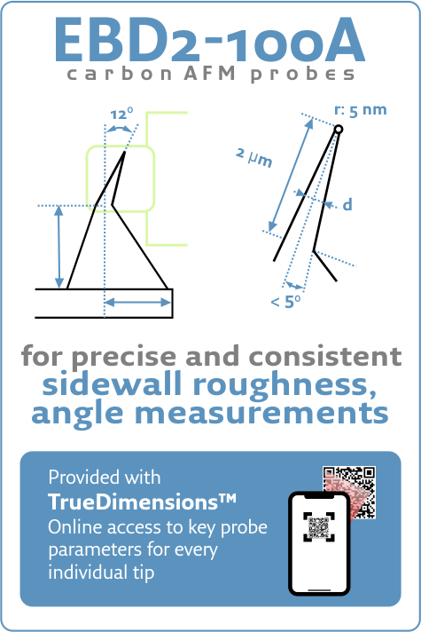
Discover how nanotools tilt compensated, high aspect ratio EBD2-100A is applied to accurately profile sidewall roughness and measure sidewall angles.
- Title: High-quality single crystal diamond diffraction gratings fabricated by crystallographic etching
DOI: 10.1364/OE.27.030371 - Authors: Marcell Kiss, Teodoro Graziosi, Adrien Toros, Toralf Scharf, Christian Santschi, Olivier J. F. Martin, and Niels Quack
- Publication: Optics Express
- Publisher: Optica Publishing Group (formerly OSA)
- Date: October 14, 2019
Real-time tracking of ionic nano-domains under shear flowMon Jun 20 2022
The behaviour of ions at solid–liquid interfaces underpins countless phenomena, from the conduction of nervous impulses to charge transfer in solar cells. In most cases, ions do not operate as isolated entities, but in conjunction with neighbouring ions and the surrounding solution. In aqueous solutions, recent studies suggest the existence of group dynamics through water-mediated clusters but results allowing direct tracking of ionic domains with atomic precision are scarce.
Read more...

Stress- and Time-Dependent Formation of Self-Lubricating In Situ Carbon (SLIC) Films on Catalytically-Active Noble AlloysThu Jun 16 2022
Although catalysis is a popular explanation for tribopolymer generation, the interplay of catalysis, mechanochemistry, and electrostatic interactions remain incompletely understood. There is consensus, however, that the mechanisms for forming a frictional polymer in situ require at least three conditions: the presence of organics, a catalytically-active substrate, and shear between surfaces (i.e., sliding contacts).
Continue reading...

BudgetSensors® ContDLC AFM Probes used in a recent studyMon Jun 13 2022
Friction force measurements with our diamond-like carbon coated ContDLC AFM probes reveal multiple symmetries for frictional anisotropy of atomic scale ripples in molybdenum disulfide
Studying MOCVD deposition of MoTe2 thin films on 8-inch SiO2/Si substratesThu Jun 09 2022
Discover how nanotools EBD-NCH with consistent radius are applied for characterizing growth, morphology, and thickness of MoTe2 thin films.
- Title: Wafer-Scale Epitaxial 1T′, 1T′–2H Mixed, and 2H Phases MoTe2 Thin Films Grown by Metal–Organic Chemical Vapor Deposition
DOI: 10.1002/admi.201800439 - Authors: TaeWan Kim, Hyeji Park, DaeHwa Joung, et al
- Publication: Advanced Materials Interfaces
- Publisher: John Wiley and Sons
- Date: Jun 4, 2018
Highlights


AFM Probe Focus


best of the best
qp-HBC
uniqprobe™ - HeartBeat Cantilever for ScanAsyst®** and Peak Force Tapping™**
Coating:
Reflective Aluminum
Tip Shape: Circular symmetric
Tip Shape: Circular symmetric
AFM Cantilever:
F
60 kHz
C
0.5 N/m
L
115 µm
ATEC-NC
Tapping Mode AFM Probe with REAL Tip Visibility
Coating:
none
Tip Shape: Visible
Tip Shape: Visible
AFM Cantilever:
F
335 kHz
C
45 N/m
L
160 µm
TESPA
Standard Tapping Mode AFM Probe
Coating:
Reflective Aluminum
Tip Shape: Standard
Tip Shape: Standard
AFM Cantilever:
F
320 kHz
C
42 N/m
L
125 µm
OTESPA
Standard Tapping Mode AFM Probe with AFM Tip at the Very End of the AFM Cantilever
Coating:
Reflective Aluminum
Tip Shape: Optimized Positioning
Tip Shape: Optimized Positioning
AFM Cantilever:
F
300 kHz
C
26 N/m
L
160 µm