

BudgetSensors® celebrates Albert Einstein's birthdayMon Mar 14 2022
"Anyone who has never made a mistake has never tried anything new." - Albert Einstein, 14 March 1879 – ∞
#AFMProbes #AtomicForceMicroscopy #HappyBirthdayEinstein

Happy World Engineering Day for Sustainable Development!Fri Mar 04 2022
Happy World Engineering Day for Sustainable Development!
“UNESCO's General Conference proclaimed the 4 March World Engineering Day for Sustainable Development (…) to raise awareness of the role of engineering in modern life, which is essential to mitigate the impact of climate change and advance sustainable development, especially in Africa and the small island developing states (SIDS).”
24 hour live stream here:


Visualizing intracellular nanostructures of living cells by nanoendoscopy-AFMWed Feb 23 2022
FIB-milled OPUS 3XC-GG AFM probes are used for cellular 3D nanoendoscopy AFM.


It's the last day of the exhibit at the 66th Biophysical Society Conference now happening in San Francisco!Tue Feb 22 2022
It's the last day of the exhibit at the 66th Biophysical Society Conference now happening in San Francisco!
AFM & SPM Probe questions? Stop by our booth 303
We are the largest SOURCE for the Top AFM Probe Brands under 1 Roof! We’ve got you covered!

NanoAndMore USA is @BiophysicalSoc meeting this week.Mon Feb 21 2022
Got any #SPMprobesquestions? Stop by our booth 303!
We're the largest source for the top #AFMProbes brands under 1 roof


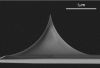
Studying Morphology, Actin Distribution and Stiffness Changes of Live Mature Epithelial CellsThu Feb 10 2022
Discover how nanotools 15 µm long, conical biotool cell XXL with 25 nm radius was applied for topographical imaging of live human cells.
- Title: Human mammary epithelial cells in a mature, stratified epithelial layer flatten and stiffen compared to single and confluent cells
DOI: 10.1016/j.bbagen.2021.129891 - Authors: Hyunsu Lee, Keith Bonin, Martin Guthold
- Publication: Biochimica et Biophysica Acta (BBA)
- Publisher: Elsevier
- Date: June 2021
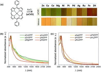
BudgetSensors® platinum coated ElectriMulti75-G AFM probes used in a recent studyThu Feb 10 2022
Our platinum coated ElectriMulti75-G AFM probes are used to measure the conductivity of oCVD coatings in this study of the impact of the central metal cation on the optoelectronic properties of directly fused porphyrin-conjugated polymers for clean and efficient solar-assisted fuel production.


Actin filaments on Mica with APTESTue Feb 08 2022
A new movie has been uploaded to the NanoAndMore AFM video gallery https://www.nanoandmore.com/video/hcggXKDrhBM
Courtesy of Prof. Noriyuki Kodera, nanoLSI, Kanazawa University, Japan you can watch a new video of #AFMprobes in action in the NanoAndMore AFM video gallery.
The video shows Actin filaments on Mica with APTES. The video was taken with #HighSpeedScanningAtomicForceMicroscopy (HS-AFM) using NanoWorld Ultra-Short #AFMCantilevers of the USC-F1.2-k0.15 type https://www.nanoandmore.com/AFM-Probe-USC-F1.2-k0.15


Akiyama-Probe (A-Probe) Motion VideoMon Feb 07 2022
Do you want to find out how the NANOSENSORS™ self-sensing and self-actuating Akiyama Probe (A-Probe) works? Then have a look at this video with its stroboscopic motion views that demonstrate how the tuning fork prongs and the AFM cantilever are really vibrating. More than 500 people have already watched it.


Happy New Lunar YearTue Feb 01 2022
We wish everyone a good start into the new lunar year of the tiger!

Happy New Lunar Year from NANOSENSORS™Mon Jan 31 2022
NANOSENSORS™ #AFMprobes wishes everyone a good start into the year of the tiger!
The NANOSENSORS screencast on the calibration service for AFM cantilevers has just reached the 1500 views mark.Sat Jan 22 2022
Accurately determined AFM cantilever properties are very important for quantitative force measurements. Force constant and resonance frequency are determined either by thermal tune, the Sader- or the dimensional method, respectively. Usually, the thermal tune method delivers the most precise values, but suffers from the fact that the AFM tip has to get in contact with the surface to calibrate the photo-detector sensitivity. This procedure may damage or break the AFM tip
Read more...
")
MikroMasch® AFM Probes used in a recent studyFri Jan 21 2022
Our AFM probes have been used in Peak Force Infrared Microscopy (PFIR), a new nano-IR technique, to analyze fine and ultrafine bioaerosols collected after filtration through a surgical mask. Some small aerosol particles below 1 micrometer can pass through facemasks.

Arrow AFM probes screencast passes 500 views markWed Jan 05 2022
The screencast about NanoWorld Arrow Silicon AFM probes held byNanoWorld AG CEO Manfred Detterbeck has just passed the 500 views mark. Congratulations Manfred!
NanoWorld Arrow™ AFM probes are designed for easy AFM tip positioning and high resolution AFM imaging and are very popular with AFM users due the highly symetric scans that are possible with these AFM probes because of their special tip shape. They fit to almost all well-known commercial SPMs (Scanning Probe Microscopes) and AFMs (Atomic Force Microscopes).

‘Live your life as an Exclamation rather than an Explanation!’Mon Dec 27 2021
‘Live your life as an Exclamation rather than an Explanation!’ Happy 379th birthday, Sir Isaac Newton!

Season’s Greetings from the whole NanoWorld® AFM probes team!Sun Dec 26 2021
Enjoy the holiday season. We're looking forward to 2022 together with you.

Happy New Year! from MikroMasch®Sat Dec 25 2021
Season’s greetings from MikroMasch®! See you again in 2022!

Season’s Greetings from NANOSENSORS AFM probesMon Dec 20 2021
There is a snowman in the park! This is a sure sign that it is again time to wish all users of our AFM probes Happy Holidays!
Take care of yourselves. We are looking forward to a new year together with you.

Max Born won the 1954 Nobel Prize in PhysicsSat Dec 11 2021
“The belief that there is only one truth and that oneself is in possession of it, seems to me the deepest root of all that is evil in the world” – Max Born, laureate of the 1954 Nobel Prize in Physics, born on 11 December 1882.
Highlights


AFM Probe Focus


best of the best
qp-HBC
uniqprobe™ - HeartBeat Cantilever for ScanAsyst®** and Peak Force Tapping™**
Coating:
Reflective Aluminum
Tip Shape: Circular symmetric
Tip Shape: Circular symmetric
AFM Cantilever:
F
60 kHz
C
0.5 N/m
L
115 µm
ATEC-NC
Tapping Mode AFM Probe with REAL Tip Visibility
Coating:
none
Tip Shape: Visible
Tip Shape: Visible
AFM Cantilever:
F
335 kHz
C
45 N/m
L
160 µm
TESPA
Standard Tapping Mode AFM Probe
Coating:
Reflective Aluminum
Tip Shape: Standard
Tip Shape: Standard
AFM Cantilever:
F
320 kHz
C
42 N/m
L
125 µm
OTESPA
Standard Tapping Mode AFM Probe with AFM Tip at the Very End of the AFM Cantilever
Coating:
Reflective Aluminum
Tip Shape: Optimized Positioning
Tip Shape: Optimized Positioning
AFM Cantilever:
F
300 kHz
C
26 N/m
L
160 µm