

The Nobel Prize in Physics 1986Fri Dec 10 2021
The Nobel Prize ceremony traditionally takes place on December 10th. The Nobel Prize in Physics 1986 was divided, one half awarded to Ernst Ruska "for his fundamental work in electron optics, and for the design of the first electron microscope", the other half jointly to Gerd Binnig and Heinrich Rohrer "for their design of the scanning tunneling microscope."


Today we remember the late Prof. Calvin QuateTue Dec 07 2021
Today we remember the late Prof. Calvin Quate, co-inventor of the Atomic Force Microscope, who was born on this date in 1923. Binnig, Quate and Gerber constructed the first AFM in 1986. The three scientists received the Kavli Prize in 2016 in recognition of their invention.


NanoAndMore USA booth no 609 at Materials Research Society MRS Fall Meeting & Exhibit 2021Fri Dec 03 2021
The annual MRS Fall is always the sign that the holiday season and and the end of the year are not far away. So with a little piece of "fan art" we would like to say thanks to all of you who visited NanoAndMore USA booth no 609 at Materials Research Society MRS Fall Meeting & Exhibit 2021. It was great to see you face to face again. Travel home safely and enjoy the holiday season.
For those of you who didn’t have the chance to visit the conference in Boston there is still the chance to visit our virtual booth from December 6 – 8, 2021 https://fallhyrbidconference.cd.pathable.com/

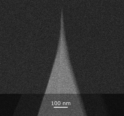
QUANTUM-AC10: Extremely Low Wear for Super-Fast AFM’sThu Dec 02 2021
Green Line product highlight:
QUANTUM-AC10: Sturdy Carbon Tip for High Speed, Fast Scanning and
Video-Rate Applications
- Consistent Sharpness
Tip radius 6-7 nm guaranteed. For reliable scanning performance and unmatched tip to tip repeatability - Controlled orientation
Tilt compensation within ±1° ensuring tip-sample perpendicularity for your specific AFM model - Scan location finder aid
Tip position mark on cantilever backside - Consistent Tuning
1.2 MHz rectangular quartz cantilevers (> 1.1 MHz guaranteed) - Diamond-like hardness and durability
Exceptional wear resistance guarantees negligible tip shape changes during high-speed scanning for consistent performance
https://www.nanotools.com/blog/quantum-ac10-extremely-low-wear-for-super-fast-afms.html
#AFM #metrology #topography #microscopy #nanotechnology

@NanoAndMore USA booth no 609 is all set up and ready to welcome youThu Dec 02 2021
@NanoAndMore USA booth no 609 is all set up and ready to welcome you @Materials_MRS MRS Fall 2021 from today until Thursday, December 2nd 2021

Last day at MRS Fall 2021Thu Dec 02 2021
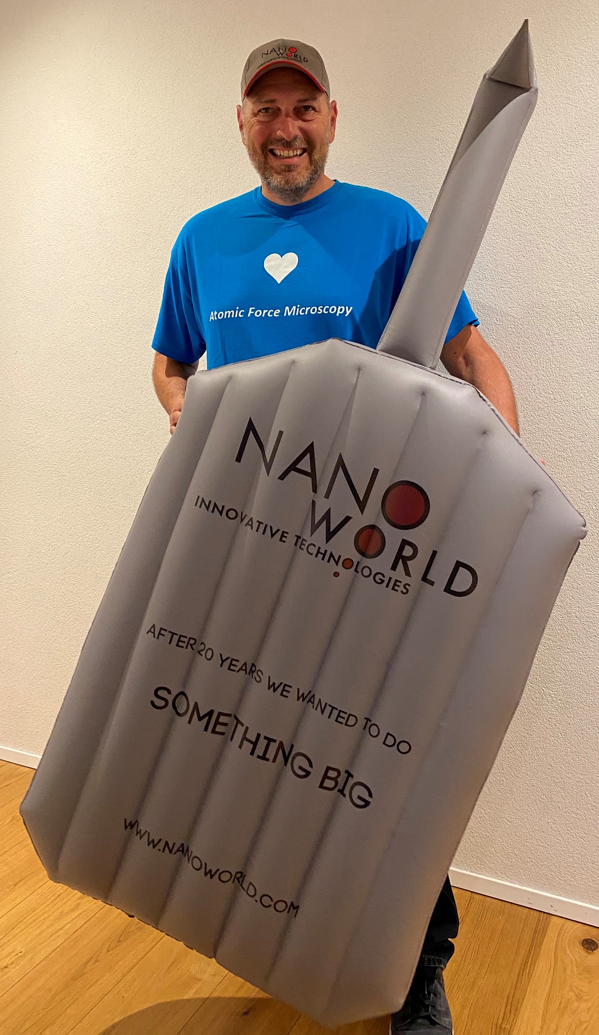
Do you need a large inflatable NanoWorld #AFMprobe for your lab or for teaching purposes? Today is your last chance to visit NanoAndMore USA booth no 609 Materials Research Society MRS Fall Meeting & Exhibit 2021 to find out how to get one of them.

Materials Research Society MRS Fall Meeting & Exhibit 2021Thu Dec 02 2021
It’s the last day at Materials Research Society MRS Fall Meeting & Exhibit 2021. NanoAndMore USA booth no. 609 will be ready for your visit from 10:00 am - 1:30 pm today.
Do you need specific #AFMprobes for your #AtomicForceMicroscopy application? Then feel free to come by and discuss your requirements with us.

Invitation to visit booth 609 at MRS Fall Exhibit 2021Wed Dec 01 2021
It’s the second day at Materials Research Society MRS Fall Exhibit 2021. NanoWorld CEO Manfred Detterbeck is at NanoAndMoreUSA booth no 609 today. We are presenting #AFMprobes in many shapes and sizes, including giant inflatable #AFMtips. Have you already visited NanoAndMore USA booth no 609 to find out more?

Feel free to pass by NanoAndMore USA booth no. 609Mon Nov 29 2021
We’re participating in the #MRS Fall Meeting & Exhibit 2021 mrs.org/fall2021 this week.
Feel free to pass by NanoAndMore USA booth no. 609 to say hello and learn more about our #AFMprobes
The exhibit hours are:
Tuesday, November 30 | 11:00 am - 5:30 pm
Wednesday, December 1 | 11:00 am - 5:30 pm
Thursday, December 2 | 10:00 am - 1:30 pm
We’re looking forward to welcoming you.

It’s the first day of the virtual Materials Research Society 2021 MRS Fall Meeting & ExhibitMon Nov 29 2021
It’s the first day of the virtual Materials Research Society 2021 MRS Fall Meeting & Exhibit
The NanoAndMore USA virtual booth is open to visitors via https://fallhyrbidconference.cd.pathable.com/
There is plenty of #AFMprobes information material available on the booth and you also have the chance to zoom with one of us in the virtual meeting room if you have any questions.

Happy Thanksgiving by NanoWorld®Wed Nov 24 2021
Happy Thanksgiving to all our friends in Canada and the USA.
Enjoy the time with friends and family safely and don’t forget to visit us at NanoAndMore USA booth no. 609 @Materials_MRS MRS Fall 2021 after the holidays if you are planning to participate in the live conference.
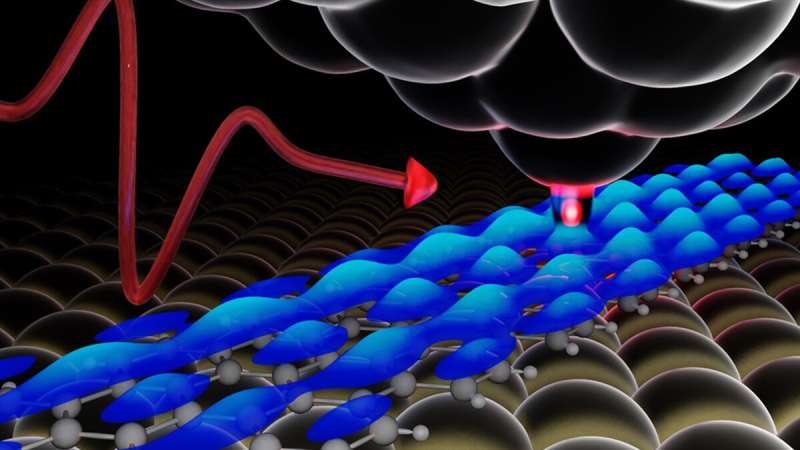
Lightwave-driven scanning tunneling spectroscopy of atomically precise graphene nanoribbonsWed Nov 24 2021
When physicist Tyler Cocker joined Michigan State University in 2018, he had a clear goal: build a powerful microscope that would be the first of its kind in the United States.
Read more...

MCNT-300™: 18 nm cylindrical tip for durable and consistent bottom access of finest featuresTue Nov 16 2021
Blue Line product highlight:
MCNT-300™: Amorphous CNT solution for demanding depth metrology applications
- Controlled shape:
Precisely controlled length of 300 nm and uniform width of 18 nm - Controlled orientation: 3° or 12°
Tilt compensation within ±0.5° for enhanced access capabilities to bottom trench features - Optimized cantilevers:
Standard with k: 40 N/m, f: 320 KHz
Softer cantilevers for force-controlling scanning modes available upon request - Diamond-like hardness and durability:
Exceptional wear resistance of diamond-like carbon for consistent
performance and reduced cost per measurement - Delivered with TrueDimensions™:
Datasheet for every single probe available online 24/7 via QR code


Happy World Science Day for Peace and Development!Wed Nov 10 2021
“Celebrated every 10 November, World Science Day for Peace and Development highlights the important role of science in society and the need to engage the wider public in debates on emerging scientific issues. It also underlines the importance and relevance of science in our daily lives.”
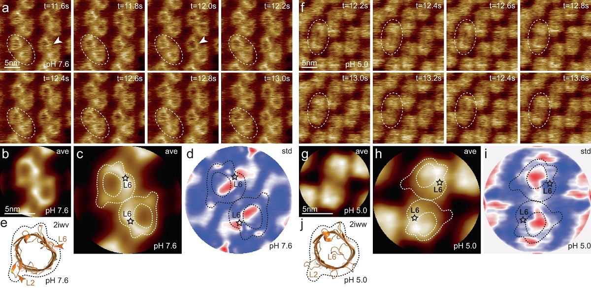
Correlation of membrane protein conformational and functional dynamicsFri Nov 05 2021
Membrane proteins (MPs) reside in the plasma membrane and perform various biological processes including ion transport, substrate transport, and signal transduction.*
Function-related conformational changes in membrane proteins occur in times scales ranging from nanoseconds to seconds.*
Continue reading...


Supercritical carbon dioxide decellularization of plant material to generate 3D biocompatible scaffoldsFri Oct 29 2021
Biocompatible scaffolds that can be repopulated with human cells have many uses such serving as replacement organs and tissues. Therefore there is an increasing interest in plant-based biomaterials for tissue engineering.*
As the above mentioned scaffolds should mimic the in vivo tissue environment closely they need to provide a fitting structural and biomechanical support to the cells while at the same time promoting cell behaviour and tissue development. *
Read more...

German National Metrology Institute (PTB) Scientists Present Nanomechanical Head with Exchangeable AFM Probes As IndenterWed Oct 20 2021
Discover how nanotools biosphere™ with precisely controlled 2 µm radius is applied to measure the mechanical properties of ultra-soft PDMS samples.
- Title: A MEMS nanoindenter with an integrated AFM cantilever gripper for nanomechanical characterization of compliant materials
DOI: 10.1088/1361-6528/ab88ed - Authors: Z Li, S Gao, U Brand, K Hiller and H Wolff
- Publication: Nanotechnology
- Publisher: IOP Publishing
- Date: 11 May 2020

MikroMasch® shared a video - how to fabricate a photo on a silicon wafer using nanotechnologySat Oct 09 2021
Check out this cool educational video from MIT.nano demonstrating how to fabricate a photo on a silicon wafer using nanotechnology.
Happy American Nanotechnology day!

BudgetSensors® celebrates Niels Bohr birthdayThu Oct 07 2021
"How wonderful that we have met with a paradox. Now we have some hope of making progress.” Today we celebrate the birthday of the late Niels Bohr, a Danish physicist famous for, among other things, developing the Bohr model of the atom.
Highlights


AFM Probe Focus


best of the best
qp-HBC
uniqprobe™ - HeartBeat Cantilever for ScanAsyst®** and Peak Force Tapping™**
Coating:
Reflective Aluminum
Tip Shape: Circular symmetric
Tip Shape: Circular symmetric
AFM Cantilever:
F
60 kHz
C
0.5 N/m
L
115 µm
ATEC-NC
Tapping Mode AFM Probe with REAL Tip Visibility
Coating:
none
Tip Shape: Visible
Tip Shape: Visible
AFM Cantilever:
F
335 kHz
C
45 N/m
L
160 µm
TESPA
Standard Tapping Mode AFM Probe
Coating:
Reflective Aluminum
Tip Shape: Standard
Tip Shape: Standard
AFM Cantilever:
F
320 kHz
C
42 N/m
L
125 µm
OTESPA
Standard Tapping Mode AFM Probe with AFM Tip at the Very End of the AFM Cantilever
Coating:
Reflective Aluminum
Tip Shape: Optimized Positioning
Tip Shape: Optimized Positioning
AFM Cantilever:
F
300 kHz
C
26 N/m
L
160 µm