NanoAndMore USA does not ship to your country
Highly efficient carbon-dot-based photoinitiating systems for 3D-VAT printingThu Jun 27 2024
Highly efficient carbon-dot-based photoinitiating systems for 3D-VAT printing
Known as a rising star among carbon nanomaterials, carbon dots (CDs) have attracted considerable interest in various fields in recent years.* In the article “Highly efficient carbon dot-based photoinitiating systems for 3D-VAT printing” Dominika Krok, Wiktoria Tomal, Alexander J. Knight, Alexander I. Tartakovskii, Nicholas T. H. Farr, Wiktor Kasprzyk and Joanna Ortyl describe how they synthesized different types of carbon dots (CDs) based on citric acid as a precursor using an efficient procedure to purify these materials from low molecular by-products and fluorophores.* They introduce three types of CDs: citric acid-based, as well as ammonia- and ethylenediamine-doped CDs, and compare […]
#3DPrintingApplications, #AFMProbes, #AFMカンチレバー, #AFMプローブ, #AFM探针, #ArrowAFMProbe, #ArrowEFM, #AtomicForceMicroscopy, #CarbonDots, #CarbonDotsCDs, #CarbonNanomaterials, #Characterization, #ConductiveAFMTip, #HydrogelPrintouts, #Hydrogels, #MaterialsResearch, #MaterialsScience, #NanoFTIR, #Nanomaterials, #NanoscaleFourierTransformInfraredSpectroscopy, #PhotoinitiatingSystems, #PolymerChemistry, #Polymers, #SSNOM, #ScanningNearFieldMicroscopy, #ScatteringTypeScanningNearFieldMicroscopy, #ScatteringTypeScanningNearFieldOpticalMicroscopy, #SurfaceCharacterization, #ナノスケールフーリエ変換赤外分光法, #原子力显微镜, #原子力显微镜探针, #散乱型走査型近接場光学顕微鏡, #散射型扫描近场光学显微镜, #电AFM探针, #纳米级傅里叶变换红外光谱
Studying the Interaction of Engineered Liposomes with Living CellsTue Jun 25 2024
Discover how nanotools biosphere™ with 2µm radius are applied to analyze adhesion forces and Young’s modulus in HeLa cells.
- Title: On the uptake of cationic liposomes by cells: From changes in elasticity to internalization
- DOI: 10.1016/j.colsurfb.2022.112968
- Authors: Adrià Botet-Carreras, Manel Bosch Marimon, Ruben Millan-Solsona, Eva Aubets, Carlos J. Ciudad, Véronique Noé, M. Teresa Montero, Òscar Domènech, Jordi H. Borrell
- Publication: Colloids and Surfaces B: Biointerfaces
- Publisher: Elsevier
- Date: January 2023
NanoAndMore Japan will be exhibiting at the 21st International Congress of BiophysicsMon Jun 24 2024
NanoAndMore Japan will be exhibiting at the 21st International Congress of Biophysics at booth number 7. International Union of Pure and Applied Biophysics
June 24 - 28, 2024, Kyoto International Conference Center https://iupab.org/event/iupabcongress2024/
Please feel free to stop by.
The huge AFM cantilever is a landmark!
Find out more about the about the big and the small #AFMprobes that we offer.
NanoAndMore Japanは、第21回国際生物物理学会議にブース番号7にて出展いたしますMon Jun 24 2024
NanoAndMore Japanは、第21回国際生物物理学会議にブース番号7にて出展いたします。@IUPAB2 https://iupab.org/event/iupabcongress2024/
2024年6月24日~28日 国立京都国際会館
お気軽にお立ち寄りください。
巨大なAFMカンチレバーが目印!
当社が提供する大型および小型 AFM プローブについて詳しくは、こちらをご覧ください。
Join us at the Panel Discussion on SPM Cantilevers at SPM Connect todayTue Jun 18 2024
Join us at the Panel Discussion on SPM Cantilevers at SPM Connect today
It’s the second day at #SPMConnect in Washington DC. There is a Panel Discussion on #SPMCantilevers today at 3:30PM – National Harbor 5. Our colleagues Manfred Detterbeck (NanoWorld CEO) and Dr. Oliver Krause (NanoWorld R&D team) will be there. Come and join us! We are looking forward to your questions and an interesting discussion.
#AFM, #AFMCantilevers, #AFMProbes, #AfmTips, #AFMカンチレバー, #AFMプローブ, #AFM探针, #AtomicForceMicroscopy, #Biophysics, #HighspeedAFM, #HSAFM, #Materialsresearch, #Materialsscience, #Nanotechnology, #PhysicalBiology, #ScanningForceMicroscopy, #ScanningProbeMicroscopy, #SPM, #SPMProbes, #SPMTips, #SPMカンチレバー, #SPMプローブ, #SPM探针, #原子力显微镜, #原子力显微镜探针, #扫描探针显微镜, #扫描探针显微镜探针
NanoAndMore USA booth at this week's SPMConnect in Washington DC from June 17-19, 2024Mon Jun 17 2024
All set up and ready to welcome you at the NanoAndMore USA booth at this week's #SPMConnect in Washington DC from June 17-19, 2024. https://www.techconnectworld.com/World2…/sym/SPMConnect.html
We're looking forward to hearing about your #scanningprobemicroscopy and #atomicforcemicroscopy applications and to many interesting talks.
Distinct Contact Guidance Mechanisms in Single Endothelial Cells and in MonolayersThu Jun 13 2024
In many #tissues, #cellshape and orientation are controlled by a combination of internal and external #biophysicalcues. *
Anisotropic #substratetopography is an ubiquitous cue that leads to cellular elongation and alignment, a process termed #contactguidance whose underlying mechanisms remain incompletely understood. *
In medium and large #arteries, atherosclerotic lesions initiate preferentially near branches and bifurcations where the #endothelialcells (ECs) lining the blood vessels are cuboidal and randomly oriented. *
In contrast, zones where endothelial cells ECs are elongated and aligned in the direction of blood flow tend to be protected from the disease. *
These observations suggest a prominent shape-function relationship and motivate interest in elucidating the mechanisms governing the regulation of EC morphology and alignment. *
In their article “Distinct Contact Guidance Mechanisms in Single Endothelial Cells and in Monolayers” Claire Leclech, Apoorvaa Krishnamurthy, Laurent Muller and Abdul I. Barakat seek to fill some of the remaining gaps in the understanding of endothelial cells shape regulation by microgrooves.
The authors try to find out, whether contact guidance responses are similar in single cells and in cellular monolayers.
To this end Leclech et al. used microgrooved substrates to investigate the contact guidance response of #vascularendothelialcells (ECs) at densities ranging from single cells at an early stage of culture as well at a later stage when the cells attain confluence, form monolayers, and exhibit collective behavior. *
The fact that the authors follow the contact guidance response of ECs in time, from #singlecells to the formation and maturation of #monolayers is the main novelty of their article. *
By tracking EC shape on #microgrooves in time and with increasing cell density, Claire Leclech et al. found that #cellalignment and elongation progressively decrease, which they interpret as a gradual loss of response to substrate topography.*
The authors could show that contrary to common belief, focal adhesion (FAs) are not the principal elements involved in the prominent groove depth-dependent contact guidance in single ECs.*
Interestingly, the contact guidance response is greatly attenuated in confluent monolayers, and cell shape and alignment in that case are driven by the organization of the #basementmembrane (BM) secreted by the cells, which leads to a loss of cellular interaction with the microgrooves.
For the characterization of the secreted basment membrane the #surfacetopography and #stiffness of the different samples were determined by #Atomicforcemicroscopy ( #AFM) with an AFM mounted on a fluorescent microscope. *
NANOSENSORS uniqprobe qp-BioAC-CI #AFMprobes with circular symmetric rounded #AFMtips with a typical tip radius of 30 nm were used.
The full citation and a direct link to the full article can be found in the NANOSENSORS blog:
A beginner’s guide to the Characterization of Hydrogel Microarchitecture for Cellular ApplicationsThu Jun 06 2024
A beginner’s guide to the #Characterization of #Hydrogel #Microarchitecture for Cellular Applications
Hydrogel materials show a number of properties which makes them interesting candidates to be utilized to mimic the #extracellularmatrix (ECM). Therefore, these materials are attractive for use in biological applications such as tissue engineering, cell culture 3D bioprinting and more.
Are you planning to use #hydrogels for the first time in your research?
Then have a look at the insightful article “A beginner’s guide to the Characterization of Hydrogel Microarchitecture for Cellular Applications” by Francisco Drusso Martinez-Garcia, Tony Fischer, Alexander Hayn, Claudia Tanja Mierke, Janette Kay Burgess and Martin Conrad Harmsen.
In their article the authors describe and evaluate the different technologies that are most commonly used to assess hydrogel microarchitecture.
Francisco Drusso Martinez-Garcia et al. explain the working principle of the various methods and also discuss the merits and limitations of each of them in view of their usefulness for the characterization of hydrogels.
They introduce and explore the pros and cons of the following methods: #ScanningElectronMicroscopy (SEM), #CryogenicScanningElectronMicroscopy (Cryo-SEM), #EnvironmentalScanningElectronMicroscopy (ESEM), #MicroComputedTomography (µ-CT), #ConfocalLaserScanningMicroscopy (CLSM), #SecondHarmonicGeneration and #AtomicForceMicroscopy (AFM).*
Atomic force microscopy ( #AFM) can be used to investigate the hydrogel #surfacetopology as well as a hydrogel’s #mechanicalproperties. The latter can be achieved through mathematical modelling of #forcedistancecurves.
When using the AFM to characterize the elasticity of a hydrogel sample it is essential to take the stiffness of the investigated material into account when choosing what kind of #AFMprobe to use for these experiments.
If an #AFMcantilever used for probing a soft sample is too stiff (if the force constant/spring constant is too high) this might result in a poor signal-to-noise ratio.
If a soft AFM probe (an AFM probe with an AFM cantilever with a low force constant) is chosen to investigate a soft material this should lead to a better signal-to-noise ratio. On the other hand, if an AFM cantilever is too soft (if the force constant is too low) then it might not be stiff enough to indent the investigated material.
Another critical factor is the shape and the size of the AFM tip.
Spheroidal AFM probes might stick to the material, resulting in artefacts, disrupted force–distance curves, or even damaged AFM cantilevers. If the #AFMtip is much smaller than the pore size of the hydrogel, it might get stuck in the fibrous network microarchitecture.
On the other hand, if the spherical AFM tip, e.g. as in #colloidalAFMprobes (a sphere glued to end of a tipless AFM cantilever), is too large, the weight of the sphere can have a negative influence on the spring characteristics of the AFM cantilever.
All these factors and more as described in the cited article have to be carefully weighed before deciding on the settings of the atomic force microscope and choosing an AFM probe for the investigation of a specific hydrogel.
@NanoWorld tipless ArrowTL2 cantilever arrays with polystyrene beads glued to them were used by the authors of this beginner’s guide to achieve the AFM data presented in the article.*
Please have a look at the NanoWorld blog for the full citation and a direct link to the full article.
Room-temperature sub-100 nm Néel-type skyrmions in non-stoichiometric van der Waals ferromagnet Fe3-xGaTe2 with ultrafast laser writabilityWed May 29 2024
The small size and magnetoelectronic properties of #magneticskyrmions make them promising candidates as data carriers for future high-density and fast-speed data storage, quantum and neuromorphic computation. *
Because of their unique atomic layered structure, two-dimensional (2D) van der Waals (vdW) #ferromagnets have some advantages when using them for practical applications in spintronic devices compared to 3D bulk ferromagnets. *
Therefore, the study of magnetic #skyrmions with small sizes, especially at room temperature (RT), in 2D vdW #ferromagneticmaterials with easy tunability has become a focal point of magnetic and topological order of matters, as well as spintronic applications. *
In the article “Room-temperature sub-100 nm Néel-type skyrmions in non-stoichiometric van der Waals ferromagnet Fe3-xGaTe2 with ultrafast laser writability” Zefang Li, Huai Zhang, Guanqi Li, Jiangteng Guo, Qingping Wang, Ying Deng, Yue Hu, Xuange Hu, Can Liu, Minghui Qin, Xi Shen, Richeng Yu, Xingsen Gao, Zhimin Liao, Junming Liu, Zhipeng Hou, Yimei Zhu and Xuewen describe how they discovered that the iron deficiency in Fe3GaTe2 can lead to a pronounced displacement of the Fe atoms within the crystal structure.*
Based on systematic structural analysis and first-principles calculations, Zefang Li et al. find that this #atomicdisplacement causes a transformation from the original centrosymmetric crystal structure to a non-centrosymmetric structure, resulting in significant Dzyaloshinskii–Moriya interactions (DMI). *
They report the discovery of a field-free sub-100 nm #Néeltypeskyrmion state in non-stoichiometric Fe2.84±0.05GaTe2 over a broad temperature range from 330 K to 100 K. *
Lorentz transmission electron microscopy (LTEM) (along with along with #magneticforcemicroscopy ( #MFM ) shows the size of the skyrmions decreases with sample thickness, and a field-free sub-100 nm skyrmion state was achieved at RT within a specific sample thickness range of 40 nm to 60 nm. *
Magnetic force microscopy (MFM), which is sensitive to the out-of-plane magnetic field in the sample, was used by Zefang Li et al. to study the effect of #nanoflake thickness on the stabilization of the #skyrmionphase at room temperature after field-cooling manipulation. *
All the nanoflakes used for the magnetic force microscopy (MFM) measurements were freshly exfoliated from the same batch to ensure consistency. *
In order to minimize the influence of the #AFMtip’s magnetic field on the #domainstructures during scanning the authors used NANOSENSORS PPP-LM-MFMR low-moment magnetic #AFMprobes.
These #MFMprobes are designed for magnetic force microscopy with reduced disturbance of the magnetic sample by the AFM tip and enhanced lateral resolution – compared to the standard MFM probes .*
The MFM measurements were carried out in an environment continuously flushed with argon gas to ensure effective protection.*
To further enhance the practical applications of their findings, Zefang Li et al. employed a homemade in-situ optical Lorentz transmission electron microscope to demonstrate ultrafast writing of skyrmions in Fe3-xGaTe2 using a single femtosecond laser pulse.*
The article by Zefang Li et al. demonstrates not only the non-stoichiometric Fe2.84±0.05GaTe2 to be a promising material platform for exploring magnetic skyrmions, but also that fs-laser can be a powerful tool to manipulate and control topological chiral spin textures to realize skyrmion-based high-speed logic and memory applications.*
Please have a look at the NANOSENSORS blog for the full citation and a direct link to the full article.
Flexible Polyurethane Foams Modified with Novel Coconut Monoglycerides-Based Polyester PolyolsFri May 24 2024
Flexible Polyurethane Foams Modified with Novel Coconut Monoglycerides-Based Polyester Polyols
The products of the polyurethane (PU) industry such as foams, coatings and adhesives are numerous and can be found in many areas of everyday life. * Polyols are an essential component in the production of polyurethane. Nowadays they mostly come from petroleum products. * In view of potential risk factors such as the running out of fossil fuels, supply chain issues, environmental concerns and economic risks it is important to develop alternatives as substitutes and supplements to the existing petroleum derived polyols. * Vegetable oils can be used to manufacture biobased polyols and various oils such as linseed oil, rapeseed […]
#AFMCantilevers, #AFMCharacterization, #AFMProbes, #AfmTips, #AFMプローブ, #AFM探针, #AtomicForceMicroscopy, #BiobasedPolyols, #CoconutOil, #FlexiblePolyurethaneFoamFPUF, #Foams, #Lipids, #MaterialsResearch, #MaterialsScience, #NCHR, #NonContactAFM, #NonContactMode, #OrganicPolymers, #Pointprobe, #PointprobeNCHR, #Polymers, #PolyolMaterials, #Polyols, #Polyurethane, #ScanningProbeMicroscopy, #SPMTips, #SPMプローブ, #SPM探针, #SustainablePolyolRawMaterial, #TappingMode, #VegetableOilBasedMaterials, #原子力显微镜, #原子力显微镜探针
Understanding Cardiac Dysfunction in Arrhythmogenic CardiomyopathyThu May 23 2024
- Title: SARS-CoV-2 protein Nsp1 alters actomyosin cytoskeleton and phenocopies arrhythmogenic cardiomyopathy-related PKP2 mutant
DOI: 10.1101/2020.09.14.296178 - Authors: Cristina Márquez-López, Marta Roche-Molina, Nieves García-Quintáns, Silvia Sacristán, David Siniscalco, Andrés González-Guerra, Emilio Camafeita, Mariya Lytvyn, María I. Guillen, David Sanz-Rosa, Daniel Martín-Pérez, Cristina Sánchez-Ramos, Ricardo García, Juan A. Bernal
- Publication: bioRxiv
- Publisher: Cold Spring Harbor Laboratory
- Date: September 16, 2020
An AFM-based approach for quantification of guest particle deformation during mechano-fusionTue May 21 2024
Research supported by our ContAl-G AFM probes presents an AFM-based approach for quantification of guest particle deformation during mechano-fusion for dry particle coatings
Lipoplex-Functionalized Thin-Film Surface Coating Based on Extracellular Matrix Components as Local Gene Delivery System to Control Osteogenic Stem Cell DifferentiationThu May 16 2024
The development of smart multifunctional #biomaterials with the ability to control the behavior of #stemcells on demand has become a powerful strategy in regenerative medicine and cell therapies.*
Stem cell-based therapies for example can offer possibilities to regenerate critical size bone defects from severe fractures or bone tissue loss after surgery. *
Various studies have determined characteristics and modifications of biomaterials that enable initiation of stem cell osteogenesis and represent promising approaches for clinical use. These approaches include materials that can mimic the bone microenvironment, materials with specific mechanical properties which stimulate bone tissue formation, and materials which can release or control the activity of osteoinductive growth factors. *
A straightforward strategy for the #functionalization of biomaterials involves emulating the properties of the #extracellularmatrix (ECM) for the formation of an artificial microenvironment that enables a precise control of cell behavior and function. *
Due to interaction of ECM components with cell surface receptors such as integrins, ECM regulates cell proliferation, migration, and differentiation.*
Therefore, the development of #surfacecoatings mimicking the native ECM structure and function is of considerable interest to functionalize implant materials. *
The #layerbylayer (LbL) technology, well-known from the pioneering work by Decher et al. on the development of #polyelectrolytemultilayers (PEMs) on solid surfaces by alternating deposition of oppositely charged polyelectrolytes, has evolved into a very simple and cost effective yet highly versatile and efficient surface modification and functionalization technology.*
The LbL technique allows the production of multifunctional thin film coatings with precise control of the film composition, structure, properties, and functions at the nanoscale. *
A further advantage of LbL is that it can be performed by different methods such as dip-coating, spray coating, and spin-coating protocols allowing the coating of different materials and designs also in a time-saving manner.*
Indeed, polyelectrolyte multilayers (PEMs) have been broadly used as reservoir for either the surface immobilization or encapsulation of bioactive molecules, more precisely drugs and proteins, to engineer bio-functional materials by choice of polyelectrolytes and complexation conditions for regenerative medicine strategies.*
Previous studies have consistently demonstrated that electrostatic-driven LbL assembly is a powerful and simple technique to functionalize biomaterials with nucleic acids aiming for non-viral gene delivery. *
In the article “Lipoplex-Functionalized Thin-Film Surface Coating Based on Extracellular Matrix Components as Local Gene Delivery System to Control Osteogenic Stem Cell Differentiation” Catharina Husteden, Yazmin A. Brito Barrera, Sophia Tegtmeyer, João Borges, Julia Giselbrecht, Matthias Menzel, Andreas Langner, João F. Mano, Christian E. H. Schmelzer, Christian Wölk and Thomas Groth describe how they combined both approaches, such as the ECM-mimicking character of PEM and their ability to be used as carrier for in situ transfection, to develop a gene-activated ECM-mimicking surface coating to direct stem cells’ fate.*
They focused on a bone ECM-mimicking PEMs consisting of Col and Cs loaded with LPX composed of OO4/DOPE lipid composite. *
Furthermore, they studied the ability of the system to induce osteogenic stem cell differentiation by gene expression analysis and mineralization assays. *
The authors present a new approach to engineer a bone-ECM inspired gene-activated surface coating which allows controlling stem cells function, and consequently, represents a promising tool to develop multifunctional surface coatings for regenerative medicine strategies. *
#AtomicForceMicroscopy ( #AFM ) was used to study the #topography and the #mechanicalproperties of the different polyelectrolyte multilayers (PEMs). *
In order to investigate the #elasticity and the #forcecurve, the samples were compressed by the #AFMtip and the #forcemapping mode was applied while the AFM tip scanned a specific area of the sample. *
The #forcemappingmode measured the #interactionforces such as adhesion or electrostatics and gives an idea regarding the stiffness and topography. The interest in testing mechanical properties and topography of surface coatings is related to their effect on #cellbehavior, such as spreading, proliferation, and differentiation. *
Atomic force microscopy with a commercially available AFM instrument in combination with an inverted fluorescence microscope was performed in #quantitativeimaging mode (QITM1) to investigate the #surfaceroughness and topography as well as record corresponding fluorescence images. *
The topographical images were recorded using @NANOSENSORS uniqprobe qp-BioT #AFMcantilevers in a standard liquid cell containing 0.15 m NaCl solution. The uniqprobe qp-BioT AFM probe types with their two different triangular AFM cantilevers (Cantilever Beam 1 (CB1) typical resonant frequency 50 kHz, typical force constant 0.3 N/m, Cantilever Beam 2 (CB2): typical resonant frequency 20 kHz, typical force constant 0.08 N/m ) offer an alternative to silicon nitride #AFMprobes, with the advantage of reduced thermal drift and taller AFM tips with smaller opening angles. *
A force map area of 5 × 5 µm2 was recorded with a resolution of 512 × 512 pixel2.
Please have a look at the NANOSENSORS blog for the full citation and a direct link to the full article
¹QITM mode is a trademark of Bruker Nano GmbH
#collagenI #lipoplexes #smartbiomaterials #boneextracellularmatrix #materialsresearch #thinfilms #polymers #DNAlipidnanoparticles #tissueengineering #quantitativeimaging #forcemapping
Happy birthday, Prof. Gerber!Wed May 15 2024
Happy birthday, professor Christoph Gerber! Thank you for your contributions to the invention of the Scanning Tunnelling Microscope, the Atomic Force Microscope and Nanoscale Science as a whole!
Prof. Gerber is the co-recipient of the 2016 Kavli Prize in Nanoscience and the recipient of the 2023 World Cultural Council Albert Einstein Award for Science.
https://cantileversensors.unibas.ch/ChGerber/Welcome.html
Visit NanoWorld booth 46 at EPFL CMi MicroNanoFabrication Annual Review Meeting todayTue May 14 2024
Come and visit our booth at the EPFL CMi 2024 MicroNanoFabrication Annual Review Meeting at the SwissTech Convention Center in Lausanne/Switzerland next Tuesday May 14, 2024 to learn more about our high-quality AFM probes. We’re looking forward to seeing you.
Correlation between plant cell wall stiffening and root extension arrest phenotype in combined abiotic stress of Fe and AlTue May 07 2024
The plasticity and growth of plant #cellwalls (CWs) is still not sufficiently understood on its molecular level. *
#AtomicForceMicroscopy (AFM) has been shown to be a powerful tool to measure the #stiffness of #planttissues. *
In the article “Correlation between plant cell wall stiffening and root extension arrest phenotype in the combined abiotic stress of Fe and Al” Harinderbir Kaur, Jean-Marie Teulon, Christian Godon, Thierry Desnos, Shu-wen W. Chen and Jean-Luc Pellequer describe the use of atomic force microscopy (AFM) to observe elastic responses of the root transition zone of 4-day-old Arabidopsis thaliana wild-type and almt1-mutant seedlings grown under Fe or Al stresses. *
In order to evaluate the relationship between root extension and root cell wall #elasticity, the authors used Atomic Force Microscopy to perform vertical indentations on surfaces of living plant roots. *
@NanoWorld Pyrex-Nitride silicon-nitride PNP-TR #AFMprobes with triangular AFM cantilevers were used for the #nanoindentation experiments with atomic force microscopy. (PNP-TR AFM cantilever beam 2 (CB2) with a typical force constant of 0.08 N/m and a typical resonant frequency of 17 kHz, typical AFM tip radius 10 nm, macroscopic half cone angles 35°). *
#Forcedistancecurves were measured using the AFM and the PNP-TR #AFMtips.*
Because of the heterogeneity of seedling CW surfaces, Harinderbir Kaur et al. used the recently developed #trimechanics3PCS framework for interpreting force-distance curves. The trimechanics-3PCS framework allows the extraction of both stiffness and elasticity along the depth of indentation and permits the investigation of the variation of stiffness with varied depth for biomaterials of heterogeneous elasticity responding to an external force. *
As shown in the article the presence of single metal species Fe2+ or Al3+ at 10 μM exerts no noticeable effect on the root growth compared with the control conditions. On the contrary, a mix of both the metal ions produced a strong root-extension arrest concomitant with significant increase of CW stiffness. *
The authors’ results indicate that the combination of Fe2+and Al3+ with exuded malate is crucial for both CW stiffening and root-extension arrest. *
It is shown that the elasticity of plant CW is sensitive and can be used to assess abiotic stresses on plant growth and stiffening. *
Please have a look at the NanoWorld blog for the full citation and a direct link to the full article.
Transparent electrodes based on molybdenum-titanium-oxide with increased water stability for use as hole-transport/hole-injection componentsSat May 04 2024
The application of #molybdenumoxide layers in electronic devices like #solarcells and #organiclightemittingdiodes (LEDs) has expanded considerably. *
Dielectric/metal/dielectric ( #DMD ) #transparentelectrodes based on #MoO3 have been applied in solar cells and organic light emitting diodes, by virtue of the favourable properties of MoO3 as hole-transport/hole-injection material.*
However, it was reported that poorly textured or amorphous MoO3 layers are extremely instable when exposed to ambient humidity and liquid water. *
In the article “Transparent electrodes based on molybdenum–titanium–oxide with increased water stability for use as hole-transport/hole-injection components” Selina Goetz, Rachmat Adhi Wibowo, Martin Bauch, Neha Bansal, Giovanni Ligorio, Emil List-Kratochvil, Christian Linke, Enrico Franzke , Jörg Winkler, Markus Valtiner and Theodoros Dimopoulos describe their study aimed to increase the water stability of sputtered, amorphous MoO3 layers without applying high temperatures, but by alloying with another refractory metal oxide to reduce hydrolysis and dissolution, while maintaining the essential electronic properties (i.e. wide band gap and high work function) of MoO3.*
In order to achieve this aim, the authors introduced titanium oxide to form a mixed molybdenum–titanium–oxide compound material. *
In contrast to the previous reports, the article by S. Goetz et al. introduces for the first time compact, amorphous #thinfilms, deposited by #sputtering from a mixed molybdenum–titanium–oxide (MTO) compound target. The sub-oxidic composition of the sputter target allows fast deposition in direct current (DC) magnetron mode, achieving high sputtering rates that are relevant for #industrialapplications. *
Selina Goetz et al. demonstrate that the addition of Ti strongly increases the stability in water, while the desirable electronic properties of MoO3, specifically the high work function and wide bandgap, are maintained. *
The DMD electrodes, with Ag as metal layer, were fabricated on both rigid and flexible substrates, namely glass and polyethylene terephthalate (PET). *
The obtained electrodes have low sheet resistance around 5 Ω/sq and high average visible transmittance well above 0.7 (including the substrate). As a result of the MTO stability, processing with water-based solutions takes place without electrode degradation. *
To demonstrate the process compatibility for large-scale, industrial production, the DMDs were sputter-deposited by a roll-to-roll process on a 300 mm-wide PET foil, achieving similar electrode properties with the laboratory-scale samples. *
The #surfacecharacterization by #atomicforcemicroscopy was performed with a commercially available #atomicforcemicroscope using NANOSENSORS SuperSharpSilicon™ SSS-NCHR silicon high-resolution #AFMprobes for tapping mode with a typical #AFMtip radius of 2 nm.
Please have a look at the NANOSENSORS blog for the full citation and a direct link to the full article.
Bi2Se3 interlayer treatments affecting the Y3Fe5O12 (YIG) platinum spin Seebeck effectThu May 02 2024
Bi2Se3 interlayer treatments affecting the Y3Fe5O12 (YIG) platinum spin Seebeck effect
Spin Seebeck effects (SSE) arise from spin current (magnon) generation from within ferri-, ferro-, or anti-ferromagnetic materials driven by an applied temperature gradient. *
Longitudinal spin Seebeck effect (LSSE) investigations, where the spin current and temperature gradient evolve along a common z axis, while the magnetic field is applied in the y axis and the voltage contacts are spaced along the x axis, have become the most popular spin Seebeck device architecture. *
In article “Bi2Se3 interlayer treatments affecting the Y3Fe5O12 (YIG) platinum spin Seebeck effect”, Yaoyang Hu, Michael P. Weir, H. Jessica Pereira, Oliver J. Amin, Jem Pitcairn, Matthew J. Cliffe, Andrew W. Rushforth, Gunta Kunakova, Kiryl Niherysh, Vladimir Korolkov, James Kertfoot, Oleg Makarovsky and Simon Woodward present a method to enhance the longitudinal spin Seebeck effect at platinum/yttrium iron garnet (Pt/YIG) interfaces. *
The introduction of a partial interlayer of bismuth selenide (Bi2Se3, 2.5% surface coverage) interfaces significantly increases (by ∼380%–690%) the spin Seebeck coefficient over equivalent Pt/YIG control devices. *
Optimal devices are prepared by transferring Bi2Se3 nanoribbons, prepared under anaerobic conditions, onto the YIG (111) chips followed by rapid over-coating with Pt. The deposited Pt/Bi2Se3 nanoribbon/YIG assembly is characterized by scanning electron microscope. The expected elemental compositions of Bi2Se3 and YIG are confirmed by energy dispersive x-ray analysis. *
A spin Seebeck coefficient of 0.34–0.62 μV/K for Pt/Bi2Se3/YIG is attained for the authors’ devices, compared to just 0.09 μV/K for Pt/YIG controls at a 12 K thermal gradient and a magnetic field swept from −50 to +50 mT. *
Superconducting quantum interference device magnetometer studies indicate that the magnetic moment of Pt/Bi2Se3/YIG treated chips is increased by ∼4% vs control Pt/YIG chips (i.e., a significant increase vs the ±0.06% chip mass reproducibility). *
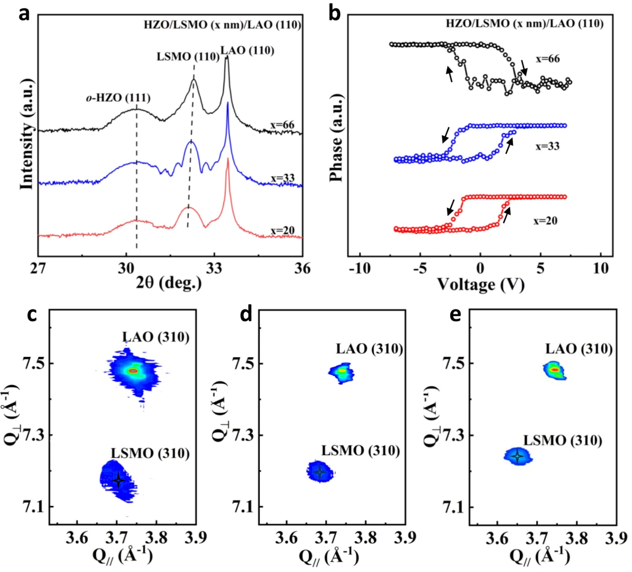
Increased surface magnetization is also detected in magnetic force microscope studies of Pt/Bi2Se3/YIG, suggesting that the enhancement of spin injection is associated with the presence of Bi2Se3 nanoribbons. *
To understand the surface magnetization effects in sample BSYIG1-a further, magnetic force microscope (MFM) measurements were undertaken using a commercial atomic force microscope and magnetic NanoWorld Pointprobe® MFMR AFM probes. *
MFM differs from traditional atomic force microscopy in that the AFM probe, in addition to providing a surface height profile, is also able to detect the magnetic field gradient above the sample. *
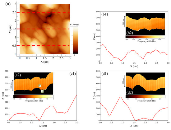
MFM surface profiling of BSYIG1-a revealed that a typical ribbon is comprised of multilayers of Bi2Se3, providing thicker sections ca. 250 nm thick [e.g., the profile along vector 1 in Figs. 3(a) and 3(b) cited below] and additional thinner sections ca. 100 nm thick [e.g., the profile along vector 2 in Figs. 3(a) and 3(b)]. Re-running ribbon profiles 1 and 2 with the magnetic probe at a height of 100 nm above the topological surface provided data on the magnetic field gradient variation along the same line profiles. The MFM amplitude [Figs. 3(c) and 3(d) cited below] increases over the Bi2Se3 flake, and furthermore, the magnetic enhancement correlates with the thickness of the Bi2Se3, being larger for the thicker part of the sample. *
This amplitude enhancement suggests that the observed effect is magnetic rather than due to long-range electrostatics, supporting the inference that the surface magnetization is improved by the presence of Bi2Se3 flakes at the interlayer of a Pt/YIG device. However, it was not possible to extract quantitative information about surface magnetization from this study, but Yaoyang Hu et al. are hopeful that future experimental and theoretical work can provide further explanation. *
Please have a look at the @NanoWorld blog for the full citation and a direct link to the full article.
Torsional force microscopy using MikroMasch® HQ:NSC18/Pt AFM ProbesFri Apr 26 2024
Torsional force microscopy using MikroMasch HQ:NSC18/Pt and other AFM probes reveals van der Waals moirés and crystal lattices in atomically thin materials.
Accurate and rapid antibiotic susceptibility testing using a machine learning assisted nanomotion technology platformWed Apr 24 2024
#Antimicrobialresistance ( #AMR) has become a significant threat to public health worldwide. *
#Nanomotion technology is based on #atomicforcemicroscopy ( #AFM) and has been proposed as potential means of circumventing these limitations by measuring bacterial viability and response to antibiotics in a growth-independent manner. *
Here, a functionalized #AFMcantilever oscillates in response to #bacterialvibrations. Conditions that alter bacterial vibrations, such as drug exposure, modulate AFM cantilever oscillations, and these changes can be detected, measured, and outputted using an optical read-out system. *
Nanomotion technology can distinguish the differing responses of resistant and susceptible bacteria to antibiotic treatment and has already been employed to detect antibiotic susceptibility for several different microorganisms. *
In the article “Accurate and rapid antibiotic susceptibility testing using a machine learning-assisted nanomotion technology platform” Alexander Sturm, et al. describe a rapid growth-independent phenotypic #antibioticsusceptibilitytests ( #AST) that uses a nanomotion technology platform to measure bacterial vibrations. *
Nanomotion-based AST strategies utilize technologies that are well-established in atomic-force microscopy (AFM). The nanomotion detection system described in the article is based on an AFM setup for cantilever-based optical deflection detection. *
However, in contrast to standard AFM devices, in this device the light source and the photodetector are placed below the AFM cantilever to facilitate the experimental workflow. A light beam, focused at the AFM cantilever end, originates in a superluminescent diode (SLED) module (wavelength: 650 mm, optical power: 2 mW), is reflected, and reaches a four-sectional position-sensitive photodetector that is a part of a custom-made precision preamplifier. *
The flexural deflection of the AFM cantilever is transformed into an electrical signal, which is further processed by a custom-made dedicated electronic module and recorded using a data acquisition card. The device is controlled using a dedicated AST software. *
The custom-made tipless AFM probes used for the described experiments are quartz-like tipless cantilevers with a gold coating acting as a mirror for the light beam (NANOSENSORS™ uniqprobe SD-qp-CONT-TL, typical spring constant: 0.1 N/m, AFM cantilever length × width × thickness: 130 × 40 × 0.75 µm, typical resonant frequency in air: 32 kHz from the NANOSENSORS Special Developments List). *
During an AST experiment, bacterial #nanoscale movements actuate the AFM cantilever to deflect in specific frequencies and amplitudes. *
The study shows the potential of this nanomotion platform for future bacterial phenotype delineation. *
Please have a look at the NANOSENSORS blog for the full citation and a direct link to the full article.
https://www.nanosensors.com/blog/accurate-and-rapid-antibiotic-susceptibility-testing-using-a-machine-learning-assisted-nanomotion-technology-platform/