Life Science AFM Probes
Atomic Force Microscopy (AFM) is a very powerful tool for studies in life sciences and biology.
On one hand AFM can measure three-dimensional topography structures with super resolution, which highlights the use of AFM for the imaging of single molecules or even single atoms. On the other hand, AFM technique can also investigate micromechanical properties of biological systems and many surface properties, such as friction, adhesion forces, viscoelastic or electrostatic properties, determining Youngs modulus or measuring interaction forces. AFM technique can obtain details of nanostructures and biomechanical properties of biological samples and analyse any kind of samples such as polymers, tissues, hydrogels, adsorbed molecules and even living cells in their native state.
But, the most important advantage of AFM for working on biological samples is the ability to operate in liquid environments, especially in buffer solutions without any extensive sample preparation. Thus, AFM is able to perform non-destructive imaging of biological material or living matter under physiological conditions. There is nearly no limitation in the choice of the medium, either aqueous or non-aqueous, sample temperature or chemical composition of the sample.
The field of applications of AFM in life sciences is huge and still growing. The investigation of cells with AFM techniques permits e.g. the characterization of their surface morphology, cytoskeletal structure, adhesion, mechanical and viscoelastic properties. Various AFM applications in mammalian cell studies have been applied e.g. in cancer cell research. Another important field of application of AFM is the study of cell membranes, especially membrane proteins and the interactions between molecules. AFM is also the only method for direct imaging and mechanical probing of liquid phase structure in a liquid environment down to the nanometer scale, which makes it a highly important tool for studying lipid membranes. AFM can also be used for structural studies of DNA or investigating DNA-protein complexes and interactions.
Besides imaging, AFM can be used as a strain gauge for studying the stretching of single molecules or fibres and as a nanomanipulator for biological systems or for the dissection of biological particles such as viruses or DNA strands. Furthermore, by linking a sensor molecule to an AFM tip one can use it as a biosensor by which single target molecules can be detected and localized on a sample surface. AFM tip modification and functionalization permit the study of specific interactions on a molecular level, such as ligand-receptor pairs (e.g. antigen-antibody-pairs), forces between molecules, cells or cell-cell interactions or intramolecular forces.
For covering as many potential applications as possible a wide range of AFM probes with different mechanical properties and made from different materials is offered by the manufacturers NANOSENSORS™, NanoWorld®, Nanotools, BudgetSensors®, MikroMasch® and OPUS by µmasch®.
The most common AFM probe materials are silicon and silicon nitride, which are available with different force constants and resonance frequencies for contact or dynamic mode imaging in air or liquid environments. The gold coating on these AFM probes enhances the reflectivity of the laser detection beam and allows applications of the AFM probes in aggressive media. AFM tip side gold coating further enables e.g. the chemical functionalization of the AFM tip using Sulphur chemistry.
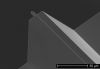
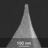
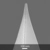
Not only AFM tip or AFM cantilever material, but also AFM tip shape is of great importance when investigating biological samples. For example, high aspect ratio AFM probes are capable of probing samples with high topography. Nanotools biotool series features precisely shaped AFM tips with hydrophobic surface properties. With an AFM tip length up to 15 µm the accessibility to subjacent regions on thick samples is provided and is combined with high resolution due to AFM tip sharpness down to 2 nm and a soft AFM cantilever for non-destructive high-resolution measurements on soft biological samples.
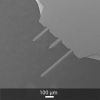
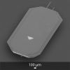
Most of the time, optimal resolution on biomolecules requires the minimum possible AFM tip radius. Nevertheless, it is sometimes preferable to use deliberately dulled AFM tips (rounded) rather than sharp AFM tips, when imaging cells for example, because the pressure exerted on the sample is reduced whereas a sharp AFM tip can poke through the membrane and damage the cell. NANOSENSORS™ qp-BioAC-CI AFM probe has a dedicated rounded AFM tip for such applications. Nano-indentation studies or probing mechanical properties of fragile biological samples may also need rounded or controlled spherical AFM tips, such as featured by the AFM tips of nanotools biosphere series or NANOSENSORS™ Special Development Sphere AFM tips.
AFM measurements in liquid environments or with temperature changes are especially sensitive to AFM cantilever drift. NANOSENSORS™ uniqprobe series is particularly adapted for such measurements as it features stress free AFM cantilevers with reduced thermal drift. Furthermore, the NANOSENSORS™ uniqprobe series due to its very small variation in force constant values from AFM probe to AFM probe is also particularly adapted to quantitative nano-mechanical studies, where a large number of AFM probes with known and near identical force constants or resonance frequencies are needed.
NanoWorld® ultra short cantilevers (USC) and Arrow ultra high frequency (UHF) AFM cantilevers can be used for real-time investigations and study of dynamic changes of biological systems. For example, dynamics of membrane components, observation of enzyme activity, mapping of dynamic mechanical properties of complex samples or monitoring microbes in real time are feasible with high-speed AFM techniques.
AFM can also be combined with different complementary techniques, including conventional optical or confocal microscopy and spectroscopy, patch clamp techniques or the use of fluidic devices to characterize a multitude of mechanical, functional and morphological properties and responses of complex biological systems.
Sort by:
106 results

PPP-BSI
Soft Contact AFM Probe for Biological Applications
Coating:
none
Tip Shape: Standard
Tip Shape: Standard
AFM Cantilever:
F
28 kHz
C
0.1 N/m
L
225 µm
ATEC-FMAu
Gold Coated Force Modulation AFM Probe with REAL Tip Visibility
Coating:
Gold Overall
Tip Shape: Visible
Tip Shape: Visible
AFM Cantilever:
F
85 kHz
C
2.8 N/m
L
240 µm
PPP-FMAu
Gold Coated Force Modulation AFM Probe
Coating:
Gold Overall
Tip Shape: Standard
Tip Shape: Standard
AFM Cantilever:
F
75 kHz
C
2.8 N/m
L
225 µm

PNP-TR-Au
Gold Coated Silicon Nitride AFM Probe
Coating:
Gold Overall
Tip Shape: Pyramid
Tip Shape: Pyramid
AFM Cantilevers: 2
1
2
F
67 kHz
17 kHz
C
0.32 N/m
0.08 N/m
L
100 µm
200 µm
USC-F1.5-k0.6
Ultra-Short Cantilever (USC) mainly dedicated to High-Speed AFM applications in liquid
Coating:
Reflective Gold
Tip Shape: Cone Shaped,EBD
Tip Shape: Cone Shaped,EBD
AFM Cantilever:
F
1500 kHz
C
0.6 N/m
L
7 µm

HQ:NSC18/Cr-Au
Gold Coated Force Modulation AFM Probe
Coating:
Gold Overall
Tip Shape: Rotated
Tip Shape: Rotated
AFM Cantilever:
F
75 kHz
C
2.8 N/m
L
225 µm
HQ:NSC19/Cr-Au
Gold Coated Force Modulation AFM Probe
Coating:
Gold Overall
Tip Shape: Rotated
Tip Shape: Rotated
AFM Cantilever:
F
65 kHz
C
0.5 N/m
L
125 µm
HQ:NSC35/Cr-Au
AFM Probe with 3 Different Gold Coated Tapping Mode AFM Cantilevers
Coating:
Gold Overall
Tip Shape: Rotated
Tip Shape: Rotated
AFM Cantilevers: 3
1
2
3
F
205 kHz
300 kHz
150 kHz
C
8.9 N/m
16 N/m
5.4 N/m
L
110 µm
90 µm
130 µm
HQ:NSC36/Cr-Au
AFM Probe with 3 Different Gold Coated Force Modulation Mode AFM Cantilevers
Coating:
Gold Overall
Tip Shape: Rotated
Tip Shape: Rotated
AFM Cantilevers: 3
1
2
3
F
90 kHz
130 kHz
65 kHz
C
1 N/m
2 N/m
0.6 N/m
L
110 µm
90 µm
130 µm

3XC-GG
AFM Probe with 3 Different Gold Coated AFM Cantilevers for Various Applications and AFM Tips at the Very End of the AFM Cantilevers
Coating:
Gold Overall
Tip Shape: Optimized Positioning
Tip Shape: Optimized Positioning
AFM Cantilevers: 3
1
2
3
F
17 kHz
150 kHz
75 kHz
C
0.3 N/m
9 N/m
2.5 N/m
L
500 µm
175 µm
240 µm
4XC-GG
AFM Probe with 4 Different Gold Coated AFM Cantilevers with AFM Tips at the Very End of the AFM Cantilevers
Coating:
Gold Overall
Tip Shape: Optimized Positioning
Tip Shape: Optimized Positioning
AFM Cantilevers: 4
1
2
3
4
F
17 kHz
75 kHz
150 kHz
1200 kHz
C
0.3 N/m
2.5 N/m
9 N/m
100 N/m
L
500 µm
240 µm
175 µm
65 µm
240AC-GG
Gold Coated Force Modulation AFM Probe with AFM Tip at the Very End of the AFM Cantilever
Coating:
Gold Overall
Tip Shape: Optimized Positioning
Tip Shape: Optimized Positioning
AFM Cantilever:
F
70 kHz
C
2 N/m
L
240 µm
XNC12/Cr-Au
AFM Probe with 2 Different Gold Coated Silicon Nitride AFM Cantilevers
Coating:
Gold Overall
Tip Shape: Pyramid
Tip Shape: Pyramid
AFM Cantilevers: 2
1
2
F
17 kHz
67 kHz
C
0.08 N/m
0.32 N/m
L
200 µm
100 µm

Multi75GB-G
Gold Coated Force Modulation AFM Probe
Coating:
Gold Overall
Tip Shape: Rotated
Tip Shape: Rotated
AFM Cantilever:
F
75 kHz
C
3 N/m
L
225 µm
ATEC-CONTAu
Gold Coated Contact Mode AFM Probe with REAL Tip Visibility
Coating:
Gold Overall
Tip Shape: Visible
Tip Shape: Visible
AFM Cantilever:
F
15 kHz
C
0.2 N/m
L
450 µm
PPP-CONTAu
Gold Coated Contact Mode AFM Probe
Coating:
Gold Overall
Tip Shape: Standard
Tip Shape: Standard
AFM Cantilever:
F
13 kHz
C
0.2 N/m
L
450 µm
PPP-NCSTAu
Gold Coated Soft Tapping Mode AFM Probe
Coating:
Gold Overall
Tip Shape: Standard
Tip Shape: Standard
AFM Cantilever:
F
160 kHz
C
7.4 N/m
L
150 µm
ContGB-G
Gold Coated Contact Mode AFM Probe
Coating:
Gold Overall
Tip Shape: Rotated
Tip Shape: Rotated
AFM Cantilever:
F
13 kHz
C
0.2 N/m
L
450 µm
HQ:CSC17/Cr-Au
Gold Coated Contact Mode AFM Probe
Coating:
Gold Overall
Tip Shape: Rotated
Tip Shape: Rotated
AFM Cantilever:
F
13 kHz
C
0.18 N/m
L
450 µm
HQ:CSC37/Cr-Au
AFM Probe with 3 Different Gold Coated Contact Mode AFM Cantilevers
Coating:
Gold Overall
Tip Shape: Rotated
Tip Shape: Rotated
AFM Cantilevers: 3
1
2
3
F
40 kHz
20 kHz
30 kHz
C
0.8 N/m
0.3 N/m
0.4 N/m
L
250 µm
350 µm
300 µm